Potato virus Y , one of the most important plant viruses affecting potato production. PVY infection of potato plants results in a variety of symptoms depending on the viral strain. The mildest symptoms is causing production loss, the most serious is called 'potato tuber necrotic ringspot disease' (PTNRD) which is transmitted by aphid vectors.
Saturday, 14 November 2009
Viruses in Potatoes
Potato virus Y , one of the most important plant viruses affecting potato production. PVY infection of potato plants results in a variety of symptoms depending on the viral strain. The mildest symptoms is causing production loss, the most serious is called 'potato tuber necrotic ringspot disease' (PTNRD) which is transmitted by aphid vectors.
Transmission of Plant Viruses
- Sap
- Insects
- Nematodes
Sap
Viruses can be spread by direct transfer of sap by contact of a wounded plant with a healthy one. Such contact may occur during agricultural practices, as by damage caused by tools or hands, or naturally, as by an animal feeding on the plant. Generally TMV, potato viruses and cucumber mosaic viruses are transmitted via sap.
Insects
Plant viruses need to be transmitted by a vector, most often insects such as leafhoppers. The chosen insect vector of a plant virus will often be the determining factor in that virus's host range. Plant virus are classified as non-persistent, semi-persistent and persiste depending on the different ways of transmission by insects.
Green peach aphid Myzus persicae, the vector of many plant viruses, including Potato virus Y.

Plant Viral Structures
Viruses are extremely small and can only be observed with an electron microscope.
Over 50% of known plant viruses are rod-shaped. The second most common structure amongst plant viruses are isometric particles.
Tobacco mosaic virus, the length is about 300-500 nm with a diameter 15-20 nm.
Viruses in Plants
Plant viruses are viruses affecting plants. They are less well known than bacteril and animal viruses, even though viruses were originally identified and isolated from tobacco plants. Plant viruses infect many food crops, including corn, beans, tobacco and the most popular in the world, potatoes, viruses resulting billions of dollars in losses each year. Viruses of plants are introduced into plant cells either through abrasions of the cell wall or by plant parasites such as nematodes and aphids.
The first virus to be discovered was Tobacco mosaic virus (TMV). This and other viruses cause an estimated US$60 billion loss in crop yields worldwide each year. The TMV viruses are most frequently used as research in plant molecular biology today.
There are over 2000 known viruses and about 1/4 of these known viruses cause diseases in plants. Plant viruses are grouped into 73 genera and 49 families.
Saturday, 7 November 2009
Viruses in Plots
Movies:
1) "Outbreak" - click here to read interview with Dr. Murphy
Novels:
Title Medusa : A novel from the NUMA files / Clive Cussler ; with Paul Kemprecos.
Author: Cussler, Clive.
Summary: Kurt Austin puts the NUMAA team on a case involving a hideous series of medical experiments, an extraordinarily ambitious Chinese criminal organization, and a secret new virus that threatens to set off a worldwide pandemic.
[From: http://catalogue.nlb.gov.sg]
Viral Scoop
“There is good circumstantial evidence that the specific origin of placental mammals is the result of an ancestral species being infected with an immunosuppressive proto-retrovirus. It is suggested that this immunosuppression permitted an immunological accommodation in the mother to the development of a genetically distinct individual in the placenta during a prolonged gestation!”
– pg 9, Basic Virology, Edward K. Wagner, Martinez J. Hewlett, David C. Bloom, and David Camerini
Wednesday, 4 November 2009
Viral Diseases in Honey Bees
"Honeybee hives have long provided humans with honey and beeswax. Such commercial uses have spawned a large beekeeping industry, though many species still occur in the wild."
Learn more about them at http://animals.nationalgeographic.com/animals/bugs/honeybee.html
Diseases
There are four diseases listed here:
- Sacbrood
- Chronic Bee Paralysis
- Black Queen Cell Virus
- Deformed Wing Virus
Sacbrood
Symptoms of sacbrood include partially uncapped cells scattered about the frame or capped cells that remain sealed after others have emerged. Diseased individuals inside cells will have darkened heads that curl upward, resembling a slipper inside the cell. The diseased prepupae fails to pupate and turns from pearl white to pale yellow to light brown and finally, dark brown. The skin is flaccid and the body watery. The dark brown individual becomes a wrinkled, brittle scale that is easily removed from the cells.
Researches estimate that one larva killed by the sacbrood virus contains enough virus to kill over one million larvae.
The viral infection process in not known as well what triggers the infection or how it persists.
Chronic Bee Paralysis
(Hairless black syndrome)
These viral infection symptoms appear only in adult bees.
They are
- trembling motion of wings and body
- unable to fly, tend to resort to crawling up to hive
- bloated abdomen and partially spread or dislocated wings
- lack of hair causes shiny and greasy appearance
As the name suggests, the virus infects the queen larvae. The immature larvae dies and turns black after its cell is sealed.
Tuesday, 3 November 2009
Viral Diseases in Rabbits
Rabbits are fairly hardy animals, and many health problems are the result of poor diet, lack of cleanliness or improper handling (which may result in injuries). Viral infections are not common in our house rabbits, and are more rampant in wild rabbits and hares which can be contracted by accident. Hence, the following is an introduction to two of the most known about and fatal infections known to kill our beloved lagomorphs.
RABBIT CALICIVIRUS (RCD) & MYXOMATOSIS
Some people are unaware that rabbits can and should be vaccinated. It is very important for rabbits to be vaccinated every year for 2 diseases, Myxomatosis and Viral Haemorrhagic Disease (VHD). These diseases are extremely serious and can be fatal if left unvaccinated.
Myxomatosis is a virus that causes an upper respiratory infection. The following are signs of Myxomatosis:
• Sticky Eyes Discharge
• Swelling around the Head and Face especially around the Eyes
• Secondary Infection called Pasteurella (Causes Pneumonia and Conjunctivitis)
"RCD Infected"
[Image from: http://members.iinet.net.au/~rabbit/Myxo2.jpg ]
This terrible virus is actually spread by fleas and biting insects. Vaccinations can be given to your rabbit at 6 weeks of age but they usually do not start until 12 weeks. If your rabbit is low risk they can be vaccinated every 12 months. Rabbits that are at high risk, such as those that live in rural areas, especially ones with a garden near their yard should be vaccinated every 6 months.
Vaccinations are generally given intra-dermally. This means that they are given in the skin rather than under the skin which is subcutaneously. Your rabbit's immune system will respond better by having the vaccination done this way. A small lump can form after the vaccination is given but it generally goes down after a day or so.
It's a sad fact that if your rabbit already has Myxomatosis, she/he may not survive. Myxomatosis can only be treated under intensive care and most rabbits do not survive it. Prevention is the best and most effective method of treatment.
Viral Haemorrhagic Disease (VHD) is almost 100% fatal and is an extremely painful death. This virus can cause the following symptoms in your rabbit:
• High Temperature
• Refuses to Eat
• Bleed from the Nose
• Convulsions
• Breathlessness
These two diseases seem to complement each other only too well.
They almost always fatal and death occurs rapidly, within 12 – 18 hours, from respiratory and heart failure. There is no known treatment. Vaccination should be at only one dose is required at the first time, then yearly boosters are required for continuing protection.
Rabbits should be kept in mosquito proof hutches, or indoors, especially early mornings and evening when mosquitoes are most active.
Control is the best option. Keep wild rabbits away from pet rabbits to prevent the spread of the fleas, as well as practice good insect control. Keep rabbits in mosquito proof hutches or indoors, especially early mornings and evening when mosquitoes are most active and use flea control on your rabbits (Advantage) as well as other pets in the household.
Here are the links to more information on this torturing method of death:
http://members.iinet.net.au/~rabbit/rcdfaq.htm
http://www.science.org.au/nova/001/cooke.htm
http://members.iinet.net.au/~rabbit/pam.htm
Viral Diseases in Cats (II)
Virologists classify feline immunodeficiency virus (FIV) as a lentivirus (or "slow virus"). It is also in the same family as the human AIDS virus, with a few significant differences. FIV is in the same retrovirus family as feline leukemia virus (FeLV), but the viruses differ in many ways including their shape. FIV is elongated, while FeLV is more circular. The two viruses are also quite different genetically, and the proteins that compose them are dissimilar in size and composition. The specific ways in which they cause disease differ, as well.
Saliva to blood (biting) is generally accepted as the primary source of spreading the virus, and it is unlikely (but not impossible) that cats will spread FIV by drinking or eating out of the same food dish, or by mutual grooming. It is not surprising that outdoor cats are particularly susceptible to the virus, and the best way to prevent infection with FIV virus is to ensure that your cat stays indoors only, which eliminates the possibility of contact with FIV cats. Another, less common means of transmission is from the mother cat to her kittens during gestation, during birth, or by nursing. There is comfort in the fact that not all FIV queens pass the virus on to their kittens. This phenomenon is not fully understood, but all kittens from FIV mothers should be tested for the FIV antibodies after six months.
If your cat has been diagnosed as FIV-positive, you'll want to work very closely with your veterinarian in designing a management program. For cats with no other symptoms, and otherwise generally good health, this might simply be a matter of ensuring he gets a sound diet, possibly with added vitamins, anti-oxidants, and Omega 3/ Omega 6 fatty acids, as well as prompt, aggressive treatment of infections and other conditions as they crop up. Even flea control is important, as fleas transmit a number of other parasites such as the Haemabartonella. Also, flea bites themselves can become infected, which would be a cause for concern.
Infected cats may appear normal for years. However, infection eventually leads to a state of immune deficiency that hinders the cat's ability to protect itself against other infections. The same bacteria, viruses, protozoa, and fungi that may be found in the everyday environment--where they usually do not affect healthy animals--can cause severe illness in those with weakened immune systems. These secondary infections are responsible for many of the diseases associated with FIV.
Early in the course of infection, the virus is carried to nearby lymph nodes, where it reproduces in white blood cells known as T-lymphocytes. The virus then spreads to other lymph nodes throughout the body, resulting in a generalized but usually temporary enlargement of the lymph nodes, often accompanied by fever. This stage of infection may pass unnoticed unless the lymph nodes are greatly enlarged. An infected cat's health may deteriorate progressively or be characterized by recurrent illness interspersed with periods of relative health.
Sometimes not appearing for years after infection, signs of immunodeficiency can appear anywhere throughout the body. Poor coat condition and persistent fever with a loss of appetite are commonly seen. Inflammation of the gums (gingivitis) and mouth (stomatitis) and chronic or recurrent infections of the skin, urinary bladder, and upper respiratory tract are often present. Persistent diarrhea can also be a problem, as can a variety of eye conditions. Slow but progressive weight loss is common, followed by severe wasting late in the disease process. Various kinds of cancer and blood diseases are much more common in cats infected with FIV, too. In unspayed female cats, abortion of kittens or other reproductive failures have been noted. Some infected cats experience seizures, behavior changes, and other neurological disorders.
Antibody tests detect the presence of antibody in the blood of infected cats. Because few, if any, cats ever eliminate infection, the presence of antibody indicates that a cat is infected with FIV. This test can be performed by most veterinary diagnostic laboratories and also is available in kit form for use in veterinary clinics. Infected mother cats transfer FIV antibodies to nursing kittens, so kittens born to infected mothers may receive positive test results for several months after birth. However, few of these kittens actually are or will become infected. To clarify their infection status, kittens younger than six months of age receiving positive results should be retested at 60-day intervals until they are at least six months old.
A negative test result indicates that antibodies directed against FIV have not been detected, and, in most cases, this implies that the cat is not infected. Nevertheless, it takes eight to 12 weeks after infection (and sometimes even longer) before detectable levels of antibody appear, so if the test is performed during this interval, inaccurate results might be obtained. Therefore, antibody-negative cats with either an unknown or a known exposure to FIV-infected cats-such as through the bite of an unknown cat-should be retested a minimum of 60 days after their most recent exposure in order to allow adequate time for development of antibodies.
" Positive for FIV, ELISA test"
On very rare occasions, cats in the later stages of FIV infection may test negative because their immune systems are so compromised that they no longer produce detectable levels of antibody.
Polymerase chain reaction (PCR) tests are designed to detect short segments of a virus's genetic material. While antibody-based tests are ideal screening tests for infection, in certain situations (such as confirming infection in antibody-positive kittens or determining infection of cats vaccinated with antibody-producing FIV vaccines), PCR-based tests, in theory, would be superior. Although PCR testing methods offer promise and are being actively explored, at this time unacceptable numbers of false-positive and false-negative results prevent them from routinely being recommended.
The only sure way to protect cats is to prevent their exposure to the virus. Cat bites are the major way infection is transmitted, so keeping cats indoors-and away from potentially infected cats that might bite them-markedly reduces their likelihood of contracting FIV infection. For the safety of the resident cats, only infection-free cats should be adopted into a household with uninfected cats.
Vaccines to help protect against FIV infection are now available. However, not all vaccinated cats will be protected by the vaccine, so preventing exposure will remain important, even for vaccinated pets. In addition, vaccination may have an impact on future FIV test results.
It is impossible to accurately predict the life expectancy of a cat infected with FIV. With appropriate care and under ideal conditions, many infected cats will remain in apparent good health for many months or years. If your cat has already had one or more severe illnesses as a result of FIV infection, or if persistent fever and weight loss are present, a much shorter survival time can be expected.
[Above info from: http://www.vet.cornell.edu/fhc/brochures/fiv.html]
Viral Diseases in Cats
If you have fallen in love with a purring kitten instead, read on. Cats have their own set of viruses which owners should know of too.
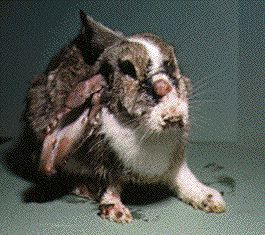
FELINE DISTEMPER/PARVOVIRUS
Feline distemper, is caused by a “parvovirus”, hence it is also known as feline parvo. It is a life-threatening disease and the virus is considered ubiquitous, meaning it is present in virtually every place that is not regularly disinfected. The infection is highly contagious among unvaccinated cats, usually kittens and young adult cats living in groups. Barn cats, feral colonies, animal shelter groups, pet stores, and rescue facilities are high risk for outbreaks. Feline distemper does not affect dogs.
The feline distemper virus amounts to a single strand of DNA surrounded by a protein coating. It is extremely stable in the environment, which leads to its characterization as “ubiquitous.” It can last a year indoors at room temperature. It survives freezing as well as treatment with such common disinfectants as alcohol and iodine. Fortunately, a 10 minute soak in bleach will kill it.
Infection occurs when the virus enters the body through the mouth or nose of the victim. Whether illness results or not depends on the immunity present in the victim vs. the number of individual virus particles entering the body.
The feline distemper virus is a “parvovirus.” Many people are familiar with this term as parvovirus infection is a very real concern for dogs, especially puppies. In fact, canine parvovirus is very closely related to the feline panleukopenia virus and much of the information regarding canine parvovirus holds true for feline distemper. The feline distemper virus, however, is more difficult to remove from the environment and more lethal in its victims than its canine counterpart.
An infected cat sheds large amounts of virus in all body secretions including faeces, vomit, urine, saliva, and mucus. The virus persists long after evidence of the original body secretion has faded away. The virus enters the victim’s body and proceeds to infect rapidly dividing cells. The lymph nodes of the throat are first and from there, over the next 2-7 days, the virus rushes to the bone marrow and intestine.
In the bone marrow, the virus suppresses production of the entire white blood cell line, hence the term “panleukopenia” (literally, “all-white-shortage”). The white blood cells are the immune cells that are needed to fight the infection and without them the victim is completely vulnerable to the advance of the virus.
In the intestine, the virus causes ulceration leading to diarrhoea and life-threatening dehydration as well as bacterial infection as the barrier between the body and intestinal bacteria is lost. The patient dies from either dehydration or secondary bacterial infection.
Because most cats are exposed to this virus to some extent, it is unusual for a kitten to have no immunity whatsoever. Further, the vaccine is so effective that even one dose can provide long lasting protection. As a result, infection is largely limited to unvaccinated younger animals kept in groups (which corresponds to exposure to amounts of virus large enough to overwhelm their partial immunity). Mortality of the sick is typically considered 90%, though it has been said that a kitten that survives the first 5 days is likely to survive the infection.
A special syndrome occurs if infection occurs during pregnancy. If infection occurs in mid or early pregnancy, the kittens simply abort. If the kittens are fairly far along, the cerebellum is involved, leading to cerebellar hypoplasia. The cerebellum is the part of one’s central nervous system that coordinates balance and movement, enabling one to walk or run on an uneven surface without consciously thinking about it. Without a normal cerebellum, the kitten is born with marked “intention tremors:” whenever he focuses on purposeful movement, he tremors so much that normal movement is impossible.
Any kitten with fever, appetite loss, diarrhoea, and/or vomiting is a suspect for feline distemper. Classically, a white blood cell count shows almost no white blood cells; there are very few causes of white cell counts this low and the infection can be considered confirmed
If a dead kitten is available for necropsy (“autopsy” in animals is called a “necropsy”), the infection is readily confirmed under the microscope as there are unique tissue findings in feline distemper.
The SNAP Fecal ELISA test kit made for canine parvovirus is often used in cats as a means to reaching a diagnosis. This test detects the presence of parvovirus in stool and is felt to be accurate though the test is not specifically labelled for this use by the manufacturer. Virus isolation, PCR testing, and antibody level measurement are also potential tests for feline distemper.
The infected cat can recover if he/she can be kept alive until his/her immune system recovers from the panleukopenia and can throw off the infection. This means that invading intestinal bacteria must be kept at bay with antibiotics and aggressive fluid therapy must control dehydration. This is essentially the same therapy as for canine parvovirus infection, though the feline experience seems to be more lethal. There is little chance of survival without hospitalization.
If a cat is lucky enough to recover from this infection, generally no permanent damage is retained and the cat goes on with lifetime immunity. The virus is shed for up to 6 weeks after recovery.
There is no way to adequately disinfect the environment; a new cat should simply be vaccinated. Vaccinations after age 12 weeks is generally effective in generating immunity against this infection, though immunity gained from mother’s milk may inactivate the vaccine through age 14-16 weeks. Maintenance boosters are generally given every 1-3 years depending on the protocol of the animal hospital. Vaccinations can be given in a nasal form or in an injection (either modified live or killed virus vaccine) given in the right shoulder area.
[Above info from: http://www.marvistavet.com/html/body_feline_distemper.html]
Viral Diseases in Dogs (III)
Canine parvovirus, or just “parvo”, is a contagious virus which affects dogs.
Two forms of CPV have been identified: diarrhoea syndrome and cardiac syndrome.
Diarrhoea syndrome, or enteritis, has an incubation period of five to fourteen days. Dogs with enteritis act like they are in extreme pain. Early symptoms are depression, loss of appetite, vomiting, high fever, and severe diarrhoea. Faeces can be either grayish or fluid and bloody. Rapid dehydration is a danger, and dogs may continue to vomit and have diarrhoea until they die, usually three days after onset of symptoms. Others may recover without complications and have no long-term problems. Puppies can die suddenly of shock as early as two days into the illness.
[

The second form of CPV is cardiac syndrome, or myocarditis, which can affect puppies under three months old. There is no diarrhoea because the virus multiplies rapidly in muscle cells of the immature heart. Puppies may stop suckling and then collapse and die within minutes or days. No effective treatment is available for cardiac syndrome, and surviving puppies may have permanently damaged hearts.
The disease is most serious in puppies, for which an untreated parvovirus has about a 90% mortality rate if it is not treated, however with treatment, the survival rate is over 80 percent. CPV infection is now considered most threatening to puppies between the time of weaning and six months of age. Adult dogs can also contract the virus, although it's relatively uncommon. All breeds of dog can be infected, but Rottweilers and Doberman Pinschers are more susceptible and have less chance of recovering.
The virus spreads through faeces – both direct and indirect contact with faeces can infect your dog. Because the virus can survive for months or even a year in soil and the environment, it is very hard to prevent any contact with the virus as it is almost impossible to clean the entire area.
Puppy mills and animal shelters are the most likely place for your dog to contract Parvovirus, due to the fact that puppies cannot have the vaccine. It is not unheard of to get a new puppy or dog from an animal shelter and find that the dog has contracted parvovirus from being in close proximity to other dogs.
The easiest way to prevent CPV in adult dogs has been through annual vaccinations, although increasingly, veterinarians are recommending that vaccinations be administered every three years. Puppies need a series of booster shots, because of uncertainty about when maternal immunity wanes and the time the vaccine can provide puppies with their own immunity. This may be as early as six weeks of age or as late as fourteen weeks of age. If there is still a high level of maternal antibody present in the puppy, it will interfere with a vaccination. Veterinarians recommend that puppies get boosters every three weeks until they are sixteen weeks old, and they should be kept separate from unvaccinated dogs. Vaccinations given to puppies as well as adults also protect against other serious canine diseases like distemper, infectious hepatitis, leptospirosis, parainfluenza, and coronavirus.
[Above info from: http://www.peteducation.com/article.cfm?c=2+1556&aid=467]
Viral Diseases in Dogs (II)
Canine distemper is one of the most significant and highly contagious viral diseases of dogs. Distemper affects multiple organs and causes numerous symptoms that increase in severity as the disease progresses. Distemper symptoms include fever, loss of appetite, eye inflammation and discharge, diarrhea, and secondary infections (e.g., pneumonia, infection of the brain and spinal cord). It is caused by a paramyxovirus, a type of virus that causes measles in humans and rinderpest in hoofed-animals such as cattle. Canine distemper affects dogs at any age. Cats, skunks, and binturong are some other animals that are also susceptible to the canine distemper virus.
Young puppies between 3 and 6 months old are most susceptible to infection and disease and are more likely to die than infected adults. Nonimmunized older dogs are also highly susceptible to infection and disease. Nonimmunized dogs that have contact with other nonimmunized dogs or with wild carnivores have a greater risk of developing canine distemper.
The canine distemper virus is spread in many ways. An infected animal can easily shed the virus through exhalation implying that the virus is transmitted via air. The virus is also shed through other bodily secretions and excretions such as urine and feces. Younger dogs are more vulnerable canine distemper than older dogs because of their under developed immune systems.
[Image from: http://www.campbowwow.com/us/il/mchenry/Portals/56/Dog%20Health/Dog%20Health%20Webpages/Distemper.jpg ]
What canine distemper is in terms of symptoms maybe confused with other diseases due to its universal and wide range clinical manifestations. Common symptoms are either gastrointestinal (such as vomiting, decrease in appetite, and diarrhea) or respiratory in nature (difficulty in breathing). Transient fever or dramatic and sporadic increases in body temperature is a telltale sign of the infection. Behavioral changes include lethargy, weakness, and depression. Neurological signs may also be seen such as muscle twitching especially near the mouth and legs. Seizures and paralysis may occur in severe cases. A more or less unique skin sign in dogs with canine distemper is the hardening of footpads and nose pad most evident in older dogs. Sudden death is not uncommon with this disease.
Aside from signs and symptoms, laboratory diagnostic examinations such as blood tests are recommended to detect and confirm the disease and its severity. Actual viral isolation and identification is also possible depending on the capacity of a laboratory.
Canine distemper is rampant in unvaccinated dog population. The distemper shot, or distemper vaccine, which often is administered with the parvovirus (also called "parvo") vaccine. Many industrialized countries having implemented great vaccination programs against the disease have successfully controlled the virus from spreading. Vaccination of young dogs begins as early as 6 weeks of age. Booster shots are administered yearly to ensure and maintain the antibodies against the virus. Basic proper hygiene and sanitation such as the use of standard disinfectants is sufficient to kill the canine distemper virus. Infected animals should also be quarantined from other animals.
Similar with other viral diseases there is no direct treatment for canine distemper. Moreover, treatment becomes irrelevant provided that preventive measures are performed. Early detection of the disease is also important to increase the chances of recovery. Once a dog has been infected, a dog owner can only offer supportive treatment and hope for the best. Antibiotic therapy may also be prescribed to lessen any detrimental effects of opportunistic secondary bacterial infections. Recovery may be absolute. However, in most cases, despite being provided with the utmost care, lingering debilitating signs do persist throughout the animal’s life such as involuntary muscle twitching.
[Above info from: http://www.animalhealthchannel.com/distemper/index.shtml]
Viral Diseases in Dogs
Now that we’ve looked at common viral diseases affecting animals, we may think that we’re safe, since we may not be farmers or people that live entirely off nature. But let’s touch on animals that are closer to our hearts, right in from our homes.
So you’ve met The One, not at the local bar, nor through a mutual friend, but on a pet farm.
You have fallen in love, bought him on your credit card and he has settled in happily with his new home. What next?
Well, you should arm yourself with some basic knowledge, because some puppies may already be infected with a virus from the pet farm, or may contract it along the way. Knowing some common ones will better prepare you on what to do once you spot the symptoms, as some of these can be fatal.
KENNEL COUGH
Dogs develop coughs too. Coughing in dogs is referred to as kennel cough and it can be a highly infectious disease. However, it is rarely serious when it strikes. Just the same, pet owners are advised to give their dogs the right amount of care and attention that they deserve whenever they are sick.
Kennel cough may give rise to a more serious health concern if not treated early on.
Kennel cough in dogs can be likened to a mild case of influenza in humans. Like influenza, the disease is also caused by a mixture of viruses and bacteria. It is a type of infection that affects the upper respiratory tract of dogs. Other household pets like cats, rabbits, and birds may develop a similar disease.
Coughs can be transferred across breeds and classes so be very careful with associating your pets when one of them is diagnosed with the disease.
More often than not, kennel cough is merely a symptom of another, much worse disease that your pet is suffering from. Kennel cough may be a result of underlying health concerns such as heart disease, periodontal disease, heartworm, parasites, throat growths, pneumonia, irritations, and allergies.
However, it is quite funny to note that some people think that kennel cough is a type of cough obtained by dogs if they live in a kennel. While dogs living in the same area may acquire the disease through association, a dog doesn't have to be inside a kennel to develop this particular type of disease.
Dogs suffering from kennel cough can be heard hacking persistently. It can be so bad at times that you might think that your dog will throw up everything it has eaten for its last meal. If you think that your dog is suffering from this disease, make sure that you take it to the veterinarian as soon as possible to keep it from suffering any longer.
Veterinarians may require inoculations or intranasal vaccines to be performed or given to your infected dog. There are also certain protective measures that you have to follow so that your dog's condition won't exacerbate. If your dog is very prone to developing kennel cough, you have to add a certain level of protection to your pet as you let it play in the park, walk it outside, or take it with you on your travels.
[Image from: http://www.basc.org.uk/filemanager/root/site_assets/howto/gundogs/administration-vaccine.jpg]
Kennel cough is considered a respiratory infection. In certain instances, antibiotics may be used as an aid to the problem. However, the choice of drugs is very crucial. Since the condition is caused by both bacteria and viruses, a double-acting antibiotic is required. Otherwise, you might to give your dog two types of oral medications, one to handle bacteria like penicillin and another to address the virus.
Treatment
There are both natural remedies and chemically prepared formulations available today to help provide relief to pets with kennel cough. However, prevention is still the best route to take. At the first signs of illness, take your dog to the veterinarian for proper diagnosis. By doing so, the symptoms of the disease will be handled accordingly and your dog will be healthier.
[Above info from: http://www.kennelcoughindogs.com/]
COMMON ANIMAL BORNE VIRUSES
Foot-and-mouth disease virus (FMDV) is the prototypic member of the Aphthovirus genus in the Picornaviridae family. This picornavirus is the etiological agent of an acute systemic vesicular disease that affects cattle worldwide. FMDV is a highly variable and transmissible virus. Soon after infection, the single stranded positive RNA that constitutes the viral genome is efficiently translated using a cap-independent mechanism driven by the internal ribosome entry site element (IRES). This process occurs concomitantly with the inhibition of cellular protein synthesis, caused by the expression of viral proteases. Processing of the viral polyprotein is achieved cotranslationally by viral encoded proteases, giving rise to the different mature viral proteins. Viral RNA as well as viral proteins interact with different components of the host cell, acting as key determinants of viral pathogenesis. In depth knowledge of the molecular basis of the viral cycle is needed to control viral pathogenesis and disease spreading.
[Image from: http://virology.wisc.edu/virusworld/images/fmdv-1qgc.jpc ]
Pestiviruses (in swine)
Pestiviruses account for important diseases in animals such as Classical swine fever (CSF) and Bovine viral diarrhea / Mucosal disease (BVD/MD). According to the current O.I.E. list CSF and BVD/MD are notifiable diseases and eradication programms are administered in many countries worldwide. The molecular biology of pestiviruses shares many similarities and peculiarities with the human hepaciviruses. Genome organisation and translation strategy are highly similar for the members of both genera. One hallmark of pestiviruses is their unique strategy to establish persistent infection during pregnancy. Persistent infection with pestiviruses often goes unnoticed; for BVDV frequently nonhomologous RNA recombination events lead to the appearance of genetically distinct viruses that are lethal to the host.
Arteriviruses (in horses)
In 1996, the family Arteriviridae was included within the order Nidovirales. Arteriviruses are small, enveloped, animal viruses with an icosahedral core containing a positive-sense RNA genome. The family includes equine arteritis virus (EAV), porcine reproductive and respiratory syndrome virus (PRRSV), lactate dehydrogenaseelevating virus (LDV) of mice and simian hemorrhagic fever virus (SHFV). Three of these viruses were first discovered and characterized a in 1964 (EAV-1953, LDV-1960 and SHFV), whereas PRRSV was first isolated in Europe and in North America in the early 1990s. The arteriviruses are highly species specific, but share many biological and molecular properties, including virion morphology, a unique set of structural proteins, genome organization and replication strategy, and the ability to establish prolonged or true persistent infection in their natural hosts. However, the epidemiology and pathogenesis of the infection caused by each virus is distinct, as are the diseases they cause.
Coronaviruses (in fowl, canines and felines)
Hendra and Nipah Virus (in swine and fruit bats)
Over the past decade, the previously unknown paramyxoviruses Hendra virus (HeV) and Nipah virus (NiV) have emerged in humans and livestock in Australia and Southeast Asia. Both viruses are contagious, highly virulent, and capable of infecting a number of mammalian species and causing potentially fatal disease. Due to the lack of a licensed vaccine or antiviral therapies, HeV and NiV are designated as biosafety level (BSL) 4 agents. The genomic structure of both viruses is that of a typical paramyxovirus. However, due to limited sequence homology and little immunological cross-reactivity with other paramyxoviruses, HeV and NiV have been classified into a new genus within the family Paramyxoviridae named Henipavirus. There is no treatment or vaccine available for either people or animals. Fruit bats of the Pteropodidae family are the natural host of Nipah virus.
Avian Influenza (in birds)

Wild aquatic birds are the natural hosts for a large variety of influenza A viruses. Occasionally viruses are transmitted from this reservoir to other species and may then cause devastating outbreaks in domestic poultry or give rise to human influenza pandemics. Proteolytic activation of the hemagglutinin is an important determinant for pathogenicity and adaptation of the receptor binding specificity of the hemagglutinin and adaptation of the polymerase to new hosts play important roles in interspecies transmission.
[Image from: http://farm1.static.flickr.com/25/61309174_35c7397efc.jpg ]
[Above info from: http://www.horizonpress.com/gateway/animal-viruses.html]
ANTIVIRAL DRUGS
Drugs that target viral processes must penetrate host cells; in doing so, they are likely to negatively impact normal pathways of the host. Antiviral drugs are characterized by a narrow therapeutic margin. Therapy is further complicated by viral latency, ie, the ability of the virus to incorporate its genome in the host genome, with clinical infection becoming evident without re-exposure to the organism. In vitro susceptibility testing must depend on cell cultures, which are expensive. More importantly, in vitro inhibitory tests do not necessarily correlate with therapeutic efficacy of antiviral drugs. Part of the discrepancy between in vitro and in vivo testing occurs because some drugs require activation (metabolism) to be effective.
Only a few agents have been found to be reasonably safe and effective against a limited number of viral diseases, and most of these have been developed in humans. Few have been studied in animals, and widespread clinical use of antiviral drugs is not common in veterinary medicine. Only a selection of the more promising agents and their purported attributes are briefly discussed.
Most antiviral drugs interfere with viral nucleic acid synthesis or regulation. Such drugs generally are nucleic acid analogs that interfere with RNA and DNA production. Other mechanisms of action include interference with viral cell binding or interruption of virus uncoating. Some viruses contain unique metabolic pathways that serve as a target of drug therapy. Drugs that simply inhibit single steps in the viral replication cycle are virustatic and only temporarily halt viral replication. Thus, optimal activity of some drugs depends on an adequate host immune response. Some antiviral drugs may enhance the immune system of the host.
Several drug classes continue to be investigated mainly because of their in vitro antiviral activities. Their potential clinical usefulness remains obscure in most instances. Included among these agents are thiosemicarbazones, guanidine, benzimidazoles, arildone, phosphonoacetic acid, rifamycins and other antibiotics, and several natural products.
[Above info from: http://www.horizonpress.com/gateway/animal-viruses.html]
Monday, 2 November 2009
How To Protect Your Children From Influenza?
When a man is diseased with influenza virus, almost all body systems are damaged including immune system, that is why recovered children feel weak and vulnerable to different viruses and bacterial infections. In order to lower disease incidence it is recommended to vaccinate not only children but also their parents.
In children from 6 months to 2 years with influenza virus, there is a high probability of complications influencing on cardiovascular and even nervous systems. According to research data, 40% of children of that age have complications with otitis, which often leads to deafness. New-born children diseases with influenza virus are subjected to complications.
The effectiveness and safety of vaccine
Vaccination practically has no contraindications but when choosing a vaccine you should consult your doctor. The effectiveness of vaccination in children is 70-100%. In children up to 6 months vaccination is less effective because of freshness of their immune system, so it is recommended to vaccinate pregnant women in second or third Trimester.
How to make a vaccine?
In order to get vaccinated you should go in one of vaccinating centers, or you can buy vaccine and use it by yourself.
Preventions of Dengue virus
The best way to reduce mosquitoes is to eliminate the places where the mosquito lays her eggs, like artificial containers that hold water in and around the home. Outdoors, clean water containers like pet and animal watering containers, flower planter dishes or cover water storage barrels. Look for standing water indoors such as in vases with fresh flowers and clean at least once a week.
The adult mosquitoes like to bite inside as well as around homes, during the day and at night when the lights are on. To protect yourself, use repellent on your skin while indoors or out. When possible, wear long sleeves and pants for additional protection. Also, make sure window and door screens are secure and without holes. If available, use air-conditioning.
If someone in your house is ill with dengue, take extra precautions to prevent mosquitoes from biting the patient and going on to bite others in the household. Sleep under a mosquito bed net, eliminate mosquitoes you find indoors and wear repellent.
[Link: http://www.cdc.gov/Dengue/prevention/index.html]
Symptoms of Dengue Virus
a) Severe headache
b) Severe eye pain (behind eyes)
c) Joint pain
d) Muscle and/or bone pain
e) Rash
f) Mild bleeding manifestation (e.g., nose or gum bleed, petechiae, or easy bruising)
g) Low white cell count
For younger children and those with their first dengue infection will have a milder illness than older children and adults.
Warning signs as temperature declines 3 to 7 days after symptoms appear:
a) Severe abdominal pain or persistent vomiting
b) Red spots or patches on the skin
c) Bleeding from nose or gums
d) Vomiting blood
e) Black, tarry stools (feces, excrement)
f) Drowsiness or irritability
g) Pale, cold, or clammy skin
h) Difficulty breathing
Go IMMEDIATELY to an emergency room or the closest health care provider
Dengue Hemorrhagic Fever
Dengue hemorrhagic fever (DHF) is characterized by a fever that lasts from 2 to 7 days, with general signs and symptoms consistent with dengue fever. When the fever declines, warning signs may develop. This marks the beginning of a 24 to 48 hour period when the smallest blood vessels become excessively permeable, allowing the fluid component to escape from the blood vessels into the peritoneum and pleural cavity. This may lead to failure of the circulatory system and shock, and possibly death without prompt, appropriate treatment. In addition, the patient with DHF has a low platelet count and hemorrhagic manifestations, tendency to bruise easily or have other types of skin hemorrhages, bleeding nose or gums, and possibly internal bleeding.
[Link: http://www.cdc.gov/Dengue/symptoms/index.html ]
Dengue Virus Vector
[Link: http://www.cdc.gov/dengue/entomologyEcology/index.html]
[Link: http://www.cdc.gov/Dengue/entomologyEcology/m_lifecycle.html]